What is fMRI?
Imaging Brain Activity
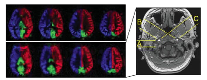
Courtesy of Dr. David Shin, UC San Diego
In your brain the activity of the neurons constantly fluctuates as you engage in different activities, from simple tasks like controlling your hand to reach out and pick up a cup of coffee to complex cognitive activities like understanding language in a conversation. The brain also has many specialized parts, so that activities involving vision, hearing, touch, language, memory, etc. have different patterns of activity. Even when you rest quietly with your eyes closed the brain is still highly active, and the patterns of activity in this resting state are thought to reveal particular networks of areas that often act together. Functional magnetic resonance imaging (fMRI) is a technique for measuring and mapping brain activity that is noninvasive and safe. It is being used in many studies to better understand how the healthy brain works, and in a growing number of studies it is being applied to understand how that normal function is disrupted in disease.
Magnetic Resonance Imaging (MRI)
MRI has become a standard tool for Radiology because it provides high resolution images with good contrast between different tissues. It works by exploiting the fact that the nucleus of a hydrogen atom behaves like a small magnet. Using the phenomenon of nuclear magnetic resonance (NMR), the hydrogen nuclei can be manipulated so that they generate a signal that can be mapped and turned into an image. When you lay in the strong magnetic field of an MRI system all of the hydrogen nuclei in your body, most of which are in water molecules, tend to align with that magnetic field. When a radio frequency (RF) magnetic pulse is applied at the right frequency, these hydrogen nuclei absorb energy and then create a brief, faint signal (the MR signal) that is detected by the RF coils in the MRI system.
The MR image is a map of the distribution of the MR signal, and by manipulating the timing of the RF pulses and the delays before detecting the signal MRI is a sensitive tool for detecting subtle changes in brain anatomy. However, mapping brain structure is not the same as mapping brain function.
fMRI

Fox MD, Raichle ME. Spontaneous fluctuations in brain activity
observed with functional magnetic resonance imaging.
Net Rev Neurosci. 2007 Sep;8(9);700-11.
The discovery that MRI could be made sensitive to brain activity, as well as brain anatomy, is only about 20 years old. The essential observation was that when neural activity increased in a particular area of the brain, the MR signal also increased by a small amount. Although this effect involves a signal change of only about 1%, it is still the basis for most of the fMRI studies done today.
In the simplest fMRI experiment a subject alternates between periods of doing a particular task and a control state, such as 30 second blocks looking at a visual stimulus alternating with 30 second blocks with eyes closed. The fMRI data is analyzed to identify brain areas in which the MR signal has a matching pattern of changes, and these areas are taken to be activated by the stimulus (in this example, the visual cortex at the back of the head).
Why is the MR Signal Sensitive to Changes in Brain Activity?
It is not because the MR signal is directly sensitive to the neural activity. Instead, the MR signal change is an indirect effect related to the changes in blood flow that follow the changes in neural activity. The picture of what happens is somewhat subtle, and depends on two effects. The first effect is that oxygen-rich blood and oxygen-poor blood have different magnetic properties related to the hemoglobin that binds oxygen in blood. This has a small effect on the MR signal, so that if the blood is more oxygenated the signal is slightly stronger. The second effect relates to an unexpected physiological phenomenon. For reasons that we still do not fully understand, neural activity triggers a much larger change in blood flow than in oxygen metabolism, and this leads to the blood being more oxygenated when neural activity increases. This somewhat paradoxical blood oxygenation level dependent (BOLD) effect is the basis for fMRI.
Blood Flow Dynamics Provides a Sensitive Window on Brain Function
Blood flow to an area of the brain is remarkably sensitive to changes in neural activity. If you sequentially tap each finger of one hand against the thumb as fast as you can, the blood flow in the motor region increases by about 60%. For this reason, blood flow changes are a sensitive indicator of underlying neural activity changes. However, these large blood flow fluctuations still result in a BOLD signal change that is only a few percent. Nevertheless, this makes it possible to map changes in activity associated with a wide range of motor, sensory and cognitive tasks. By carefully designing experiments to probe different aspects of brain function, many investigators are trying to better understand the neurological and psychological changes associated with Alzhemer’s disease, schizophrenia, depression, autism and many other disorders.
In addition, in recent years it has become clear that there is a great deal of information on how the brain is organized just in the way different brain regions continue to fluctuate together even when you are not doing a particular task. The strength of these resting state networks (RSN) also changes with disease, and an important goal is to investigate whether psychiatric disease can be understood in terms of disorders of these basic networks.
Measuring Blood Flow Directly with ASL
The BOLD signal that underlies most fMRI applications is essentially a qualitative signal, because it depends in a complex way on the combined physiological changes of blood flow and oxygen metabolism. An alternative MRI method called arterial spin labeling (ASL) is a tool for measuring blood flow changes directly. One of the limitations of the BOLD signal is that it is always a signal change between two conditions, such as tapping your fingers compared to resting. For this reason, BOLD imaging can tell us nothing about the actual level of blood flow before the task started. With ASL it is possible to measure the absolute level of blood flow in any condition. For example, if blood flow decreases as Alzheimer’s disease develops, this could be detected with ASL methods but not with BOLD imaging. ASL works by manipulating the MR signal of arterial blood before it is delivered to different areas of the brain. By subtracting two images in which the arterial blood is manipulated differently, the static signal from all the hydrogen nuclei in the rest of the tissue subtracts out, leaving just the signal arising from the delivered arterial blood.
ASL and BOLD imaging can be used together to provide a more quantitative probe of brain function, including assessment of oxygen metabolism changes, and this potential synergy is a primary motivation for ongoing research at the CFMRI in developing the next generation of fMRI methods.
Diffusion Tensor Imaging (DTI)
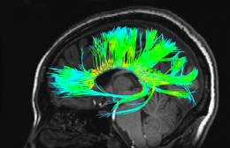
Brain function depends on the wiring between brain regions, the complex web of axons carrying signals from one neuron to another. In addition to methods for detecting brain activation with fMRI, MRI also provides a way to measure these anatomical connections. The white matter of the brain consists of bundles of these axonal fibers, so that within a small region the fibers are all aligned, and diffusion tensor imaging (DTI) is able to measure the direction of this alignment. Knowing the orientation of the fibers at each point it is possible to trace paths through the brain that map the fiber tracts. The method exploits the sensitivity of the magnetic resonance signal to the small random motions of water molecules. This diffusion of water molecules is analogous to a drop of ink slowly expanding in a pool of water as the ink molecules diffuse. In white matter fiber tracts the displacements of water molecules due to diffusion are much greater along the direction of the fibers than in a perpendicular direction, making it possible to map the fiber orientation with DTI. In addition to mapping white matter fiber tracts, these methods are useful for detecting and characterizing disorders of white matter in disease.
MR Microscopy
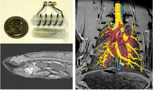
Using our 7 Tesla scanner and special purpose coils designed for high resolution imaging, it is possible to image mice (and even zebrafish) with a resolution well below one tenth of a millimeter. While many of these studies also target the brain, these studies also extend outside the brain and include detailed identification of anatomical structures (such as the airways of the mouse lung in this image) for a variety of experiments. The insights into anatomy and physiology made possible by high resolution imaging in small animals provides a critical complement to the human studies on our 3 Tesla scanners.
Structural/Anatomical MRI
Structural MRI provides information about brain anatomy to complement functional MRI in a number of ways. Since brain function depends to some extent on the integrity of brain structure, measures that characterize the underlying tissue integrity allow one to examine the impact of tissue loss or damage on functional signals. Furthermore, structural MRI provides anatomical reference for visualization of activation patterns and regions of interest to extract functional signal information.
Many pulse sequences are available, emphasizing different aspects of normal and abnormal brain tissue. By modifying sequence parameters such as repetition time (TR) and echo time (TE), for example, anatomical images can emphasize contrast between gray and white matter (e.g., T1-weighted with short TR and short TE) or between brain tissue and cerebrospinal fluid (e.g., T2-weighted with long TR and long TE).
Information from structural MRI can be used to describe the shape, size, and integrity of gray and white matter structures in the brain. Morphometric techniques measure the volume or shape of gray matter structures, such as subcortical nuclei or the hippocampus, and the volume, thickness, or surface area of the cerebral neocortex. The volume of normal and abnormal white matter also can allow inference of macrostructural white matter integrity, providing indications of inflammation, edema, and demyelination; complementary microstructural studies using diffusion imaging can help to provide a more comprehensive picture of white matter integrity.
Combining structural MRI, functional MRI and diffusion imaging may more broadly characterize normal and abnormal brain function, supporting biomarker studies of neurodegenerative or psychiatric disorders to determine risk, progression, and therapeutic effectiveness.


